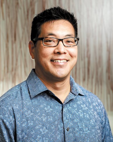
Pathologists: Lab Work, Diagnoses
Dr. Christopher Lum
Director of dermatopathology services for Hawaii Pathologists’ Laboratory at The Queen’s Medical Center
Where did you receive your schooling and training?
I did my undergraduate and graduate work at University of Hawaii in microbiology. I went to John A. Burns School of Medicine and then went away for my pathology internship and residency at Los Angeles County-University of Southern California General Hospital. From there, I completed a year in diagnostic surgical pathology fellowship at USC, before doing my subspecialty training fellowship at Cornell-Memorial Sloan Kettering in cutaneous (skin-related) pathology or dermatopathology. I also completed a molecular diagnostics certification by American Board of Clinical Chemistry. In addition to directing the dermatopathology services at Hawaii Pathologists’ Laboratory, I’m the associate medical director of HPL’s molecular diagnostics laboratory.
What got you interested in dermatopathology?
I was a technician here at Queen’s pathology lab, and I worked with a pathologist who saw a lot of skin diseases. Dermatopathology has a lot of Latin, eponyms and history, so this was interesting to me. In addition, dermatopathologists and dermatologists work closely together because you need to correlate what you’re seeing on the slide with what they’re seeing clinically on the patient. That clinical dynamic makes the slide diagnoses more real and personalized.
How long have you been working in pathology?
About 25 years. I started in 1990 as data-entry clerk, typing patient demographics into the lab report, then I moved up to being a phlebotomist and drew blood in the clinical lab. Then I was as a path-tech (pathology technologist) for a long time before going off to medical school. I’ve been practicing at HPL as a pathologist since 2005.
What happens in the pathology department?
Pathologists are in charge of lab tests. Many medical decisions that a physician makes with their patient uses some data point from the laboratory. In the surgical pathology laboratory, we receive tissue from surgical operations and surgical procedures, both in the hospital and in the clinic office. We take that tissue and look at it under a microscope to make a diagnosis whether what we’re looking at is benign or cancer. When we look at it closer we can tell whether this tumor has features of good patient survival or poor patient survival. We list those features in our pathology report.
Oncologists use the diagnostics we have gathered to judge treatment and life expectancy. Our pathology report has information that shows what stage the cancer is in. A very aggressive therapy may make sense for a stage II cancer, but in a stage IV cancer, where the ultimate outcome may be eventually death, an aggressive therapy may not be the best choice. Also clinical or experimental trials might be better in a stage IV category than in stages I or II. Our report provides that information to the patients and their families, so that they can make a better decision. With an institution like Queen’s, there are many specialists involved in cancer: Radiation therapists, surgical oncologists, medical oncologists, patient navigators, geneticists. When a malignant call is made, there are a lot of resources that will be instantly geared up to treat that.
Can you talk about the difference between clinical and surgical lab diagnostics?
There are two main branches of lab tests. There’s the blood, body fluid and microbiology branch, which is the clinical laboratory. Then, there’s the surgical pathology laboratory which is involved in looking at biopsy material and excision specimens that come from out-patient clinics and from the main OR (operating room).
Are pathologists only in the lab or do they sometimes interact with patients?
I think there’s a misconception that pathologists are hidden away in a basement closet and don’t interact with people. On the contrary, I think good pathologists have to be interactive and accessible. I try to be accessible to patients and their inquiries about their pathology reports. I’ve spoken with patients on the phone regarding my findings, explained terms, discussed literature on diseases, and some come in to look at their slides under the microscope.
There is a branch of surgical pathology, called cytology, that has a close interaction with patients. Cytology uses a technique called fine needle aspiration (FNA) biopsies. A small needle is placed into a palpable mass and the contents of the needle are spread on a glass slide. The slide is stained using the staining units on the cart, and looked at under the microscope. It’s a diagnosis right there. Our lab has an active FNA service. Our carts go out to hospital bedsides and physician offices.
We also often pair with radiology to do CT-guided and ultrasound-guided biopsies. When we look at the tissue at the bedside in the radiology suite we can let them know immediately whether they have diagnostic tissue or not. We let them know whether the mass is benign or malignant. We can also let them know whether they have enough tissue for other tests that are critical for the diagnosis. It saves the patient from having to book another appointment and another needle stick. For neighbor island patients, it saves them from flying back and forth. Studies have shown that when a pathologist goes to the bedside, the false negative rate is a lot lower. That’s good for hospital resources and for the patient.
Do pathologists do autopsies?
Yes. Hospital autopsies have gone down over the last decades, because of better imaging. Still, autopsies are good for our active residency programs. A lot of information is gained by doing an autopsy, both from the educational standpoint at the medical student level, to know anatomic structures, but also from the clinical standpoint for the medical resident who helped with treating the patient. They can see the severity and extent of disease.
Do pathologists do CSI?
Crime scene investigation belongs to a subspecialty of anatomic pathology, called forensics. We only work with medically related diseases, primarily cancer.
With dermatological pathology, do you only work with cancers, or also material swabbed from rashes or other skin conditions?
My main focus is on cancers of the skin, so melanoma is very important, squamous cell carcinoma, basal cell carcinoma, some cutaneous lymphomas of the skin. Sometimes when there is a dermatitis or rash that is problematic, the dermatologist will biopsy it and I can add additional information to help them make a decision on what it is.
Does HPL do lab work only for Queen’s?
Our pathology group covers Queen’s Hospital and its campuses, as well as the Neighbor Islands and even the Pacific Basin, all the way to Guam, American Samoa, Saipan. We’re the oldest pathology group in the islands. We celebrate our 50th anniversary this year.
Anything else you’d like to mention?
We have a summer pathology internship program geared for students who are interested in medicine. We’re in an exciting time for pathology. It’s an old science but there’s constant innovation. Now that the human genome has been sequenced, we’re able to begin using that knowledge and technology for patient care. We’re very excited to introduce our summer researchers to this new capacity in medicine.
In years prior, stage for stage, a cancer was treated the same. The unlocking of the genetic code to patient care has identified molecular targets to treat cancer. These targets are different from patient to patient, so detection of these genetic changes provides a more personalized, precise approach to cancer care. Pathologists play a critical role in detecting these alterations and directing that care.
For more information, call 547-4271.


